Friday, May 30, 2025

MRI Defecography Report Template

๐Ÿ“ Sample MRI Defecography Report Template

Patient Name:                                                                   Age/Sex:
Referring Physician:


Clinical Indication: Chronic constipation, pelvic organ prolapse, incomplete evacuation, etc.

Technique:

MRI Defecography was performed in the supine position. The rectum was distended with ~120–180 ml of ultrasound gel. Dynamic sequences were obtained at rest, during squeeze, strain, and defecation.

Findings:

1. Rectum:

  • At rest: Normal / Redundant / Dilated

  • During defecation: Normal emptying / Incomplete evacuation

  • Rectocele: Present / Absent – Size (e.g., 2.5 cm)

  • Intussusception: Yes / No – Describe level

  • Mucosal prolapse: Present / Absent

2. Anal Canal:

  • Length: ___ cm

  • Sphincter integrity: Intact / Disrupted (internal/external)

  • Anismus: Yes / No – (Failure of relaxation noted)

3. Pelvic Floor Descent:

  • Pubococcygeal Line (PCL) reference used.

  • Perineal descent: Mild / Moderate / Severe (e.g., >3 cm below PCL)

4. Anterior Compartment (Bladder/Urethra):

  • Cystocele: Yes / No – Grade (I/II/III)

  • Urethral hypermobility: Present / Absent

5. Middle Compartment (Uterus/Vagina):

  • Uterine prolapse: Yes / No

  • Vaginal vault descent: Yes / No – Degree

6. Posterior Compartment:

  • Enterocele: Yes / No – Bowel loop between vagina and rectum

Impression:

  • Findings suggest a ___ (e.g., moderate rectocele with internal intussusception and pelvic floor dyssynergia).

  • Recommend correlation with clinical symptoms and pelvic floor physiotherapy or surgical consultation.

Thursday, May 29, 2025

MRI prostate plain, and contrast, What is the Function of the Prostate?

1. What is the Function of the Prostate?

The prostate is a small gland (about the size of a walnut) located below the bladder in men and surrounds part of the urethra.

Main functions:

  • Produces seminal fluid: This fluid mixes with sperm from the testes to form semen.

  • Nourishes and protects sperm: The fluid from the prostate contains enzymes, zinc, and citric acid that support sperm function.

  • Aids ejaculation: During ejaculation, the prostate contracts to help propel semen into the urethra.

2. Why Do MRI Prostate Plain (Non-Contrast)?

Plain MRI of the prostate (also called non-contrast or multiparametric MRI) is used as a non-invasive diagnostic tool.

Reasons:

  • Evaluate prostate anatomy and size

  • Detect tumors: Especially suspicious areas that may indicate prostate cancer.

  • Assess lesions: Determine if a lesion is likely to be benign or malignant.

  • Guide biopsies: Helps target areas that need tissue sampling.

MRI prostate plain typically includes:

  • T2-weighted imaging: Excellent for viewing the prostate’s internal zones.

  • Diffusion-weighted imaging (DWI): Detects how water molecules move in tissue, helping find cancer.

  • Apparent diffusion coefficient (ADC): Quantifies DWI.

  • Sometimes, spectroscopic imaging.

Many centers prefer non-contrast multiparametric MRI (mpMRI) as the initial step for screening or evaluating prostate issues.

3. Why Do MRI Prostate With Contrast (Dynamic Contrast Enhanced MRI – DCE-MRI)?

MRI with contrast (using gadolinium-based agents) is done when:

  • Further clarification of a lesion is needed.

  • Higher suspicion of cancer is present.

  • Evaluating cancer spread (staging).

  • Checking vascularity of a lesion (cancers typically show early enhancement).

  • Monitoring after treatment (e.g., after surgery, radiotherapy).

Dynamic Contrast Enhanced (DCE) imaging helps:

  • Detect fast-enhancing areas (which may indicate aggressive tumors).

  • Improve PI-RADS scoring (used to assess prostate lesions).

Sunday, May 25, 2025

MRI Defecography MRI Defecography Protocol (Basic).

 MRI Defecography (also called MR Defecography or Dynamic Pelvic Floor MRI) is a specialized MRI study used to evaluate the structure and function of the pelvic floor muscles during defecation. It's particularly useful for patients with chronic constipation, incontinence, pelvic organ prolapse, or a sense of incomplete evacuation.

๐Ÿ” Purpose

MRI Defecography helps assess:

  • Pelvic organ prolapse (rectocele, cystocele, enterocele)

  • Rectal intussusception

  • Anismus (failure of anal sphincter to relax)

  • Pelvic floor dyssynergia

  • Perineal descent

๐Ÿงช Procedure Overview

  1. Patient Preparation:

    • Mild laxatives may be used before the exam.

    • Patients are often asked to avoid eating for a few hours prior.

  2. Intraluminal Contrast:

    • The rectum is filled with ultrasound gel or jelly to simulate stool.

    • Sometimes, the vagina or bladder is also filled for better visualization of all compartments.

  3. MRI Imaging:

    • Images are acquired at rest, squeeze, strain, and defecation phases.

    • A true sagittal plane is primarily used.

    • Additional axial and coronal planes may be used for anatomical detail.

✅ Advantages

  • Non-invasive

  • No ionizing radiation

  • Detailed soft tissue contrast

  • Multi-compartment evaluation (anterior, middle, posterior pelvic compartments)

๐Ÿ“Š Typical MRI Sequences Used

  • T2-weighted fast spin echo (static anatomy)

  • Balanced steady-state free precession (for dynamic phases)

  • Cine imaging during defecation for motion assessment

๐Ÿฉบ Clinical Indications

  • Suspected complex pelvic floor disorders

  • Failed conventional treatment for constipation or incontinence

  • Prior to surgical planning for pelvic floor repair

๐Ÿ“ท MRI Defecography Protocol (Basic)

SequencePlanePurpose
T2 FSEAxial/SagittalStatic soft tissue anatomy
Balanced SSFP (TrueFISP/FIESTA)SagittalDynamic imaging at rest, squeeze, strain, defecation
Cine MRSagittalVideo loop for functional assessment
Optional T1AxialFor detecting fat or hemorrhage

๐Ÿ“Š Infographic Idea for Radiographic Gyan

Title: "MRI Defecography: A 360° Look at the Pelvic Floor"

Sections:

  1. What is MRI Defecography?

    • Purpose and advantages

  2. How is it done?

    • Step-by-step with icons (gel injection, squeeze, strain, defecate)

  3. Anatomical Areas Assessed

    • Rectum, bladder, uterus/vagina

  4. Common Findings

    • Rectocele, intussusception, enterocele, perineal descent

  5. When is it needed?

    • Symptoms that indicate the scan

Friday, May 23, 2025

MRI sequences and its use.

 MRI (Magnetic Resonance Imaging) sequences are various techniques used in MRI scans to obtain different types of images and contrast. Each sequence is designed to highlight different tissues or conditions in the body. Here’s a rundown of some commonly used MRI sequences:

1. T1-Weighted Imaging (T1WI)

  • Purpose: Provides high-resolution images of anatomical structures.
  • Appearance: Fat appears bright; water and cerebrospinal fluid (CSF) appear dark.
  • Uses: Good for assessing anatomy and structural details. Often used post-contrast to evaluate lesions or tumors.

2. T2-Weighted Imaging (T2WI)

  • Purpose: Highlights differences in water content.
  • Appearance: Water and CSF appear bright; fat and most solid tissues appear darker.
  • Uses: Useful for identifying edema, inflammation, and many pathologies. It’s often used to assess brain lesions and spinal cord abnormalities.

3. Proton Density Imaging (PD)

  • Purpose: Measures the density of hydrogen protons in tissues.
  • Appearance: Provides contrast based on the density of protons rather than their relaxation times.
  • Uses: Good for evaluating structures where the contrast between tissues is less pronounced but still informative.

4. Fluid-Attenuated Inversion Recovery (FLAIR)

  • Purpose: Suppresses the signal from fluids (like CSF) to highlight lesions.
  • Appearance: CSF is dark, while lesions in the brain are bright.
  • Uses: Effective for identifying lesions in the brain, particularly useful in diagnosing multiple sclerosis and other demyelinating diseases.

5. Gradient Echo (GRE)

  • Purpose: Provides images with varying contrast depending on the echo time (TE) and repetition time (TR).
  • Appearance: Sensitive to magnetic field inhomogeneities.
  • Uses: Useful for imaging blood products, calcifications, and sometimes for functional MRI (fMRI).

6. Echo Planar Imaging (EPI)

  • Purpose: Fast imaging technique that acquires images quickly.
  • Appearance: High speed and can be prone to artifacts.
  • Uses: Common in functional MRI (fMRI) and diffusion tensor imaging (DTI).

7. Diffusion Weighted Imaging (DWI)

  • Purpose: Measures the diffusion of water molecules in tissues.
  • Appearance: Areas with restricted diffusion (e.g., acute stroke) appear bright.
  • Uses: Important for diagnosing strokes and assessing the integrity of white matter.

8. Diffusion Tensor Imaging (DTI)

  • Purpose: An extension of DWI that maps the diffusion of water in multiple directions.
  • Appearance: Provides information on the orientation and integrity of white matter tracts.
  • Uses: Used in research and clinical practice to study brain connectivity and white matter abnormalities.

9. Susceptibility Weighted Imaging (SWI)

  • Purpose: Enhances the visibility of blood products and calcifications.
  • Appearance: Highlights areas with different magnetic susceptibilities.
  • Uses: Effective for detecting microbleeds, venous structures, and calcifications.

10. Magnetic Resonance Angiography (MRA)

  • Purpose: Visualizes blood vessels without the need for contrast injection.
  • Appearance: Shows blood vessels as bright structures against a darker background.
  • Uses: Non-invasive assessment of vascular structures, such as arteries and veins.

Each MRI sequence has specific applications and is chosen based on what the clinician is looking to diagnose or evaluate.

Wednesday, May 21, 2025

Why Do Cardiac MRI? Preparation for Cardiac MRI, Procedure of Cardiac MRI ?


๐Ÿซ€ Why Do Cardiac MRI?

Cardiac MRI (Magnetic Resonance Imaging) is done to provide detailed images of the heart’s anatomy and function without using radiation. It helps diagnose and monitor various heart conditions more precisely than other imaging modalities like X-ray, CT, or echocardiography.

Common Reasons to Perform a Cardiac MRI:

  1. Cardiomyopathy – Detect hypertrophic, dilated, or restrictive types.

  2. Myocardial infarction – Assess damage, scarring, and tissue viability.

  3. Congenital heart disease – Detailed anatomy of structural heart defects.

  4. Myocarditis/Pericarditis – Identify inflammation or fluid.

  5. Heart tumors or masses – Characterize abnormal growths.

  6. Aortic diseases – Aneurysms or dissections.

  7. Valve problems – Analyze function and structure.

  8. Cardiac function evaluation – Ejection fraction, stroke volume, wall motion.

cardiac mri prepration
why do cardiac mri

๐Ÿงช Preparation for Cardiac MRI

1. Patient Screening:

  • Check for metal implants, pacemakers, or other MRI-incompatible devices.

  • Ask about claustrophobia or anxiety.

2. Consent & Medical History:

  • Explain the procedure and take informed consent.

  • Review kidney function if contrast (Gadolinium) is needed.

3. Fasting:

  • Usually 4–6 hours fasting if contrast or pharmacologic stress is used.

4. Clothing and Accessories:

  • Remove all metallic objects: jewelry, hairpins, hearing aids, etc.

  • Change into MRI-safe gown.

5. Medication:

  • Beta-blockers may be given to slow heart rate (for better image quality).

  • Sedation if the patient is claustrophobic (optional and under supervision).


๐Ÿ› ️ Procedure of Cardiac MRI

1. Positioning:

  • Patient lies supine (on back) on MRI table.

  • ECG leads and a chest coil are attached for cardiac gating.

2. Planning Scans:

  • Localizer images are taken to plan precise imaging planes (short axis, long axis, etc.).

3. Imaging Sequences:

  • Cine MRI: Assesses cardiac motion.

  • T1/T2 Mapping: For tissue characterization.

  • Late Gadolinium Enhancement (LGE): To detect scarring or fibrosis.

  • Perfusion Imaging: May involve stress agent to check blood flow.

4. Contrast Injection (if needed):

  • Gadolinium-based contrast injected via IV to highlight blood vessels and scar tissue.

5. Monitoring:

  • Continuous ECG and verbal communication.

  • Procedure typically lasts 30–60 minutes.

6. Post-Scan:

  • Patient observed briefly.

  • Hydration advised to flush out contrast.

Sunday, May 18, 2025

Types of Cardiac MRI.

 Cardiac MRI (Magnetic Resonance Imaging of the heart) can be classified based on the purpose of the scan, technique used, and clinical indication.

๐Ÿ”น Types of Cardiac MRI (Based on Purpose and Technique)

  1. Cine MRI

    • Shows real-time heart motion ("movie" of the beating heart).

    • Used to assess cardiac anatomy, ventricular function (ejection fraction), and wall motion.

  2. Late Gadolinium Enhancement (LGE) MRI

    • Uses contrast to detect myocardial scar, fibrosis, or infarction.

    • Common in ischemic heart disease and cardiomyopathies.

    • cardiac mri types
      types of cardiac mri


  3. Perfusion MRI

    • Assesses blood flow to the myocardium at rest and stress.

    • Used to detect ischemia (similar to a nuclear stress test).

  4. T1 and T2 Mapping

    • Quantitative imaging of myocardial tissue properties.

    • Helps in detecting edema, fibrosis, or infiltration (e.g., amyloidosis, myocarditis).

  5. MR Angiography (MRA)

    • Visualizes blood vessels including coronary arteries and great vessels.

    • Can be done with or without contrast.

  6. Phase Contrast MRI

    • Measures blood flow velocity and direction.

    • Used for assessing valvular diseases and congenital heart defects.

  7. 4D Flow MRI

    • Advanced form of phase-contrast MRI.

    • Shows flow patterns in 3D + time.

  8. Stress Cardiac MRI

    • Done with pharmacologic stress agents (e.g., adenosine or dobutamine).

    • Assesses myocardial perfusion and function under stress.


types of cardiac mri

๐Ÿ”น
Types Based on Clinical Indication

  • Ischemic Heart DiseasePerfusion, LGE, Cine MRI

  • Cardiomyopathies (e.g., HCM, DCM)Cine, LGE, T1/T2 mapping

  • Myocarditis or Infiltrative DiseaseT1/T2 mapping, LGE

  • Congenital Heart DiseaseMR Angiography, Cine, Flow MRI

  • Valvular Heart DiseaseCine, Phase Contrast MRI

  • Pericardial DiseaseCine, LGE, T2 mapping

Wednesday, May 14, 2025

Is MRI Contrast Safe During Pregnancy? can we do contrast scan in permanency patient?

Is MRI Contrast Safe During Pregnancy?

MRI with contrast (Gadolinium-based contrast agents) is generally avoided during pregnancy, unless absolutely necessary.

Why is Gadolinium Contrast Risky in Pregnancy?

  • Gadolinium crosses the placenta and enters fetal circulation.

  • It has been shown to:

    • Stay in the amniotic fluid longer.

    • Potentially accumulate in fetal tissues.

    • Animal studies have shown toxic effects at high doses.

According to the American College of Radiology (ACR) and FDA, gadolinium should only be used during pregnancy if the potential benefit clearly outweighs the potential risk to the fetus.

 Safe MRI Practice During Pregnancy

  • Non-contrast MRI is considered safe at any stage of pregnancy.

  • Contrast MRI is typically reserved for life-threatening conditions or situations where the diagnosis cannot be made without contrast (e.g., some tumors, vascular malformations).


    is mr contrast safe for preganancy
    can we do contrast scan in permanency patient?

 Summary

MRI TypeSafety in PregnancyNote
Non-contrast MRI                      SafePreferred option
Gadolinium Contrast MRI                     Use only if essential                                   Risk of fetal exposure

If MRI with contrast is needed for a pregnant patient, always ensure:

  • A radiologist and referring physician document the justification.

  • Informed consent is taken.

  • Use the lowest effective dose of a macrocyclic agent (e.g., Gadavist, Dotarem).

Saturday, May 10, 2025

GE MRI Signa HDxt MRI

 The GE Signa HDxt MRI is a widely used MRI system from GE Healthcare, available in multiple configurations, typically 1.5 Tesla. It's designed for whole-body imaging and is known for its upgradeability and image quality. Here's a detailed list of its main components and equipment.

๐Ÿงฒ System Overview: GE Signa HDxt MRI

  • Magnet Strength: 1.5 Tesla (also available in 3.0T variants)

  • Bore Size: 60 cm

  • Field of View (FOV): Up to 48 cm

  • Gradient System: TwinSpeed with 33 mT/m amplitude and 100 T/m/s slew rate (varies by model)

  • Software Platform: GE Excite or HDxt software platforms with upgrade options

  • RF Channels: Typically 8-channel or 16-channel systems (expandable in some versions)

๐Ÿงฐ Main Equipment and Components

1. Magnet Subsystem

  • Superconducting magnet

  • Cryogenic cooling system (uses helium)

  • Active/passive shielding

2. Gradient Subsystem

  • Gradient coils (X, Y, Z)

  • Gradient power amplifiers

  • Cooling system for gradient coils

3. RF System

  • Transmit RF coil (body coil inside bore)

  • Multiple receive coils (see below)

  • RF amplifiers

  • Digital RF chain (DSP, ADC)

4. Coils (Receive/Transmit)

  • Head coil (for brain imaging)

  • Neck coil

  • Spine array coil

  • Body array coil

  • Knee coil

  • Shoulder coil

  • Breast coil

  • Cardiac coil

  • Peripheral vascular coil

5. Patient Handling System

  • Patient table (motorized, detachable in some models)

  • Table controls (manual and remote)

  • Positioning aids and straps

  • Emergency release system

6. Computer and Operator Console

  • Operating console with dual monitors

  • Scan planning software

  • Image post-processing tools

  • PACS connectivity

  • DICOM compatibility

7. Cooling System

  • Helium compressor unit (cryocooler)

  • Chiller unit (external)

  • RF and gradient coil cooling circuits

8. Shielding and Room Requirements

  • RF shielded room (Faraday cage)

  • Magnetic shielding (if required)

  • Quench pipe (for helium venting)

9. Other Accessories

  • Ear protection for patients

  • Intercom system

  • Physiological monitoring system (ECG, respiratory gating)

  • Injector pump for contrast agents (optional)

๐Ÿงช Applications & Sequences

  • Neuroimaging (fMRI, DTI)

  • Musculoskeletal

  • Body imaging (Abdomen, Pelvis)

  • Cardiovascular MRI

  • Breast MRI

  • Angiography (MRA)

  • Diffusion/Perfusion imaging

  • Spectroscopy (optional)

✅ Optional Upgrades/Features

  • HD gradients (for better resolution)

  • Advanced software licenses (e.g., for cardiac or neuro applications)

  • Silent Scan (noise reduction technology)

  • GEM suite coils (higher channel count and comfort)

Wednesday, May 7, 2025

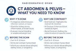
Why should you do a CT Abdomen and Pelvis WHAT CAN DETECT CONTRAST SCAN?

 1. Why should you do a CT Abdomen and Pelvis?

A CT scan of the abdomen and pelvis is performed to:

  • Investigate abdominal pain

  • Detect tumors, infections, or inflammation

  • Identify internal injuries or bleeding

  • Evaluate digestive tract conditions

  • Monitor the progress of diseases or response to treatment

  • Guide surgical planning, biopsies, or drainage procedures


2. What can be detected in a CT Abdomen and Pelvis?

It can help detect or evaluate:

  • Appendicitis

  • Diverticulitis

  • Pancreatitis

  • Kidney stones

  • Liver disease (e.g., fatty liver, cirrhosis, tumors)

  • Tumors or masses (in liver, pancreas, kidneys, intestines, etc.)

  • Enlarged lymph nodes

  • Aortic aneurysms

  • Bowel obstruction or perforation

  • Abscesses or fluid collections

  • Bladder abnormalities


WHAT CAN DETECT CONTRAST CT SCAN
WHY SHOULD DO CT ABDOMEN PELVIS


3. Why should you do an oral contrast scan?

Oral contrast is used to:

  • Highlight the gastrointestinal (GI) tract (stomach, small intestine, colon)

  • Help distinguish bowel loops from other abdominal structures

  • Detect bowel perforations, masses, or inflammation

  • Evaluate for fistulas or abscesses

4. Why is IV (intravenous) contrast needed?

IV contrast is used to:

  • Enhance visibility of blood vessels and organs

  • Differentiate between normal and abnormal tissues

  • Improve detection of tumors, infections, or vascular conditions

  • Show organ perfusion and inflammation

5. Why is rectal contrast sometimes used in CT Abdomen and Pelvis?

Rectal contrast is given to:

  • Better outline the colon and rectum

  • Evaluate for colitis, bowel perforation, or rectal tumors

  • Assess anorectal abscesses or fistulas

  • Improve visualization in post-surgical cases or complex infections

6. What should you do before a CT Abdomen and Pelvis?

  • Fasting: Don’t eat for at least 4–6 hours before the scan (especially if IV/oral contrast is used).

  • Hydration: Drink plenty of water unless advised otherwise.

  • Oral Contrast: You may be asked to drink oral contrast 1–2 hours before the scan.

  • Medical History: Inform the radiologist or technician about:

    • Allergies (especially to iodine or contrast)

    • Kidney issues

    • Diabetes (especially if on metformin)

    • Pregnancy

  • Arrive early: You may need time for prep like contrast drinking or IV setup.

Tuesday, May 6, 2025

Difference between CT scan and MRI.

 The difference between CT scan and MRI lies mainly in the technology used, the type of images produced, and their applications. Here's a clear comparison.

๐Ÿง  1. Technology Used

  • CT Scan (Computed Tomography)

    • Uses X-rays to create detailed cross-sectional images of the body.

    • Multiple X-ray images are taken from different angles and compiled by a computer.

  • MRI (Magnetic Resonance Imaging)

    • Uses strong magnetic fields and radio waves to generate images.

    • No ionizing radiation is involved.


difrance between ct scan and mri
difrance between ct scan and mri 


๐Ÿ–ผ️ 2. Image Detail and Use

FeatureCT ScanMRI
Bone ImagingExcellent for bones and fracturesNot as good as CT
Soft TissueDecent, but less detailedSuperior for soft tissues (brain, spine, joints)
SpeedFast (few minutes)Slower (15–60 minutes)
Emergency UsePreferred in trauma and strokeLess commonly used in emergencies
CancerUseful for detecting tumors and stagingExcellent for soft tissue tumor detail

3. Safety and Contraindications
  • CT Scan

    • Involves radiation, so it's used with caution, especially in children and pregnant women.

  • MRI

    • No radiation, but not suitable for patients with metal implants, pacemakers, or certain tattoos due to strong magnets.

๐Ÿ” 4. Cost

  • CT Scan is generally cheaper and more widely available.

  • MRI is more expensive and may require more time and preparation.

๐Ÿ“ Summary Table:

AspectCT Scan         MRI
Imaging Method            X-rays                   Magnetic field + radio waves
Best for            Bones, lungs, trauma cases                   Brain, muscles, spinal cord
Radiation            Yes                   No
Time            Quick                   Longer
Cost            Less expensive                   More expensive
Metal Implants            Usually safe                   Not safe

MRI Defecography Report Template

๐Ÿ“ Sample MRI Defecography Report Template Patient Name:                                                                    Age/Sex: Refer...